Class 11 Biology Ch 20 Locomotion and Movement important points to remember
Welcome student, today we will mention that Class 11 Biology Ch 20 Locomotion and Movement important points to remember, it will cover the whole NCERT, and best for revision, and prepared by the masters of their field.
Class 11 Biology Ch 20 Locomotion and Movement important points to remember
Types of Movement :
1. Amoeboid movement : These movements takes place in phagocytes where leucocytes and macrophages migrate through tissue. It is affected by pseudepodia formed by the streaming of protoplasm (as in amoeba)
2. Ciliary movement : These movement occurs in internal organs which are lined by ciliary epithelium.
3. Muscular Movement : This movements involve the muscle fibers, which have the ability to contract and relex.
Properties of Muscle : (i) Excitability (ii) Contractility
(iii) Extensibility (iv) Elasticity
Types of Muscles :
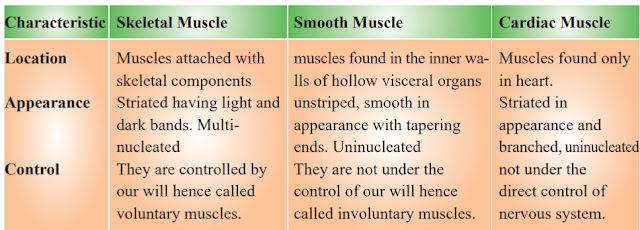
(a) Skeletal muscles or striated muscles—These involved in locomotion and change of body postures. These are also known as voluntary muscles.
(b) Visceral muscles or smooth muscles—These are located in inner wall of hollow visceral organ, smooth in appearance and their activity are not under control of voluntary nervous system. They are called involuntary muscles.
(c) Cardiac muscles—The muscles of heart, involuntary in nature, striated and branched, These are uninucleated.
Structure of myofibril :
- Each myofibril consist of alternate dark and light band.
- Dark band—contain myosin protein and is called A-band or Anisotroic band.
- Light band—Contain actin protein and is called I Band or Isotropic band.
- I Band is bisected by an elastic fiber called ‘Z’ line. Actin filament (thin filament) are firmly attached to the ‘Z’ lines.
- Myosin filament (thick filament) in the ‘A’ Band are also held together in the middle of T Band by thin fibrous membrane called ‘M’ line.
- The portion between two successive ‘Z’ lines is considered as functional unit of contraction and is called a sarcomere.
Structure of Actin and Myosin Filament
Red muscle fibres :
White muscle fibres
— These are pale or whitish due to presence of less content of myoglobin.
— These contain fewer mitochondria
— Sarcoplasmic reticulum is more/high
— During sternous exercise, lactic acid accumulates in large quantity so muscle fatigues
Mechanism or Muscle contraction : Sliding filament theory
The contraction of muscle fiber takes place by the sliding of actin (thin
filament) on myosin (thick filament)
- Muscle contraction is initiated by a signal sent by the CNS via a motor neurone.
- Impulse from motor nerve stimulates a muscle fiber at neuro muscular junctions.
- Neurotransmitter releases here which generates an action potential in sarcolema.
- This causes release of Ca++ into sarcoplasm. These Ca++ binds with troponin, thereby remove masking of active site.
- Myosin head binds to exposed active site on actin to form a cross bridge, utilising energy from ATP hydrolysis.
- This pulls the actin filament towards the centre of ‘A’ band.
- ‘Z’ lines also pulled inward thereby causing a shortening of sarcomere i.e. contraction.
- I band get reduced, whereas the ‘A’ band retain the length.
- During relexation, the cross bridge between the actin and myosin break.
Ca++ pumped back to sarcoplasmic cisternae. Actin filament slide out of ‘A’ band and length of I band increase. This returns the muscle to its original state.
Vertebral formulae of man C7T12L5S(5) C(4) = 33
Thanks for reading it, I recommend you to attempt the MCQ question of it which is uploaded by our website click below link to attempt it.


No comments
Give your thoughts